
-

联系电话
热线电话
+86-15221725700
-

添加微信
微信扫一扫添加

-

回到顶部
联系我们
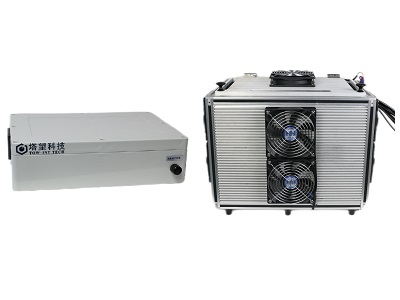
动物光照暴露系统
产品介绍
视网膜是视觉形成的重要组织,过度光照会造成视网膜的损伤,随着光学设备及仪器的普遍 使用导致视网膜光损伤也不断发生,因此视网膜光损伤是眼科领域的研究热点。在对视网膜光损伤机制进一步研究的过程中,由于临床试验开展具有一定的局限性,这就要求我们能建立拟合临床要求的视网膜光损伤动物模型。 塔望科技生产的动物光照暴露系统是专为视网膜光损伤动物建模设计,可以稳定的提供亮度可调的光暴露环境。标准提供 LED 光源,0-250k lux 可调。另外有蓝光暴露系统供选择。
技术参数
1. 用于视网膜光损伤动物建模
2. 光源:LED 光源,白色,波长 450-465nm
3. 适用动物:小鼠(可定做大鼠)
4. 光照强度最大范围:0-250K lux,建议使用范围 0-200K lux,其他亮度可定做
5. 内尺寸:40*25*25cm,可容纳小鼠笼一个,其他尺寸可定做
6. 外尺寸:50*40*40cm
7. 整机重量:25kg
8. 电源/功率:220V,1200W
9. 配备光照测量仪 1 个
参考文献
Tool from ancient pharmacopoeia prevents vision loss
Jeffrey H. Boatright,1 Anisha G. Moring,1 Clinton McElroy,1,2 Michael J. Phillips,1,2 Vi T. Do,1 Bo Chang,3 Norm L. Hawes,3 Amber P. Boyd,1 Sheree S. Sidney,1 Rachael E. Stewart,1 Steven C. Minear,1 Rajashree Chaudhury,1 Vincent T. Ciavatta,1,2 Cecilia M.P. Rodrigues,4 Clifford J. Steer,5 John M. Nickerson,1 Machelle T. Pardue1,2
1 Department of Ophthalmology, Emory University School of Medicine, Atlanta, GA; 2 Atlanta VA Medical Center, Research Service, Decatur, GA; 3 The Jackson Laboratory, Bar Harbor, ME; 4 Centro de Patogenese Molecular, Faculty of Pharmacy, University of Lisbon, Lisbon, Portugal; 5 Department of Medicine, University of Minnesota Medical School, Minneapolis, MN
Molecular Vision 2006; 12:1706-14
Fundus Camera-Delivered Light-Induced Retinal Degeneration in Mice With the RPE65 Leu450Met Variant is Associated With Oxidative Stress and Apoptosis
Xin Zhong,* Bogale Aredo, Yi Ding, Kaiyan Zhang,† Cynthia X. Zhao, and Rafael L. Ufret-Vincenty Department of Ophthalmology, University of Texas Southwestern Medical Center, Dallas, Texas, United States
IOVS j October 2016 j Vol. 57 j No. 13 j 5559
Increased susceptibility to fundus camera-delivered light induced retinal degeneration in mice deficient in oxidative stress response proteins
Yi Ding1,2,* , Bogale Aredo1,* , Xin Zhong1,3, Cynthia X. Zhao1, and Rafael L. Ufret-Vincenty1 1Department of Ophthalmology, UT Southwestern Medical Center, Dallas, Texas, 75390-9057 2Current address: Department of Ophthalmology, The Central Hospital of Wuhan, Tongji Medical College, Huazhong University of Science and Technology, Wuhan, Hubei, 430014, P.R. China 3Current address: Department of Ophthalmology, The First Affiliated Hospital of Guangxi Medical University, Nanning, Guangxi, 530021, P.R. China
Exp Eye Res. 2017 June ; 159: 58–68. doi:10.1016/j.exer.2017.03.009
Increased expression of ceruloplasmin in the retina following photic injury
Lin Chen, Tzvete Dentchev, Robert Wong, Paul Hahn, Rong Wen, Jean Bennett, Joshua L. Dunaief F. M. Kirby Center for Molecular Ophthalmology, Scheie Eye Institute, University of Pennsylvania, Philadelphia, PA
Molecular Vision 2003; 9:151-8
*我公司可提供3Q验证,根据客户的特殊应用、特殊需求提供功能定制服务,也可以提供相关的实验服务,详情请来电咨询。

 2021-08-24
 次
次